Parkin/PINK1 Mitophagy Evidence And pS65-Ub Detectability Notes
Parkin/PINK1 Mitophagy Evidence Matrix
Pathologic Row / Park2 Mutation
Row labels and effect-chain fragments:
pathologic
Park2 mutation
LOF
↓ pUb binding with Parkin (G284R, R275W)
abnormal protein folding & ↓ protein structural stability
(2019 Yi?) -> ↓ protein expression
↓ Ub? phosphorylation (R42P, V56E?) -> ↓ Parkin activation
↓ ubiquitination
↓ fission
↓ mitophagy
Mito rupture
↑ mt DNA release
↓ MC1 mass/activity
↓ ATP
↑ ROS
↑ Apoptosis
Neuronal loss
Upper-column fragment:
(mitochondria network) by Parkin's effect on DRP1Extracellular Mitochondrial Expulsion Figure
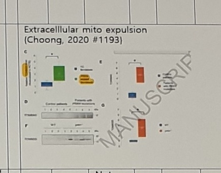
Figure title:
Extracellular mito expulsion
(Choong, 2020 #1193)
Figure labels and numeric/statistical marks are too small to transcribe safely from this photo.
Evidence Rows
Evidence-row entries from the matrix:
| Evidence type | Stable visible content |
|---|---|
| Postmortem human | 아직 못 발견; Not found |
| Human CSF | cf Parkin |
| Human serum | Parkin can be detected in CSF & serum (Castellazzi, 2019 #891) |
| Mouse | (Palacino, 2004 #726): ↓ complex I & iv, ↓ ETC capacity, ↓ mito respiration, but normal morphology; (Noda, 2020 #701) KO Mouse, fragmented mito; (Palacino, 2004 #726): ↓ proteins re protection from OX; (Noda, 2020 #701) neuronal loss (2y) |
| iPS | (Suzuki, 2017 #680) ↑ ROS in park2 iPS; (Suzuki, 2017 #680) ↑ apoptosis (cleaved caspase3-positive cells (Fig. 3B).) in park2 iPS |
| fibroblast | 아래 'fibroblast 참조' |
Biomarker And Antibody Notes
Notes in the lower part of the matrix:
include LOF
Exclude Hyperactive Parkin variants (by known sequencing? Fibroblast?)
only MC1 PET+Ve patients?

Antibodies to PINK1 are being developed by MJFF (Padmanabhan, 2019 #820)

pS65 ub,
- Can we discriminate free pS65-ub and that bound to Parkin?
- Should be increased by PARKIN GT in non-neuronal cell type
  (그렇지만 gt roa 상 말초에서 변화 측정은 무의미하겠네)

pS65 Parkin antibody is being developed by MJFF (Padmanabhan, 2019 #820)

Antibody to activated Parkin is being by MJFF (Padmanabhan, 2019 #820)

Parkin substrate: VDAC1 (2018 Ordureau)
- Antibodies to Parkin substrates (VDAC, TOMM20, MFN2) are being developed by MJFF
  (Padmanabhan, 2019 #820)

Mitophagy in peripheral?
(fibroblast?, cf, mitophagy in heart tissue in mice, 2018 Sliter fig1)

BCPP MC1 imaging

(Geldenhuys, 2014 #733):
Parkin In sPD And pS65-Ub Detectability
Parkin in sPD row:
Parkin in sPD

postmortem
(Lonskaya, 2013 #1742) 12sPD,
(↑ phosphorylation -> ) ↓ (~50%, WB) Parkin level &
↓ soluble parkin, ↑ insoluble parkin (in striatum,) (not in cortex),
SN 은 안 본 듯
pS65-Ub reference text:
expression of pS65-ub

(Fiesel, 2015 #700): The increase of pS65-Ub upon mitochondrial damage was not
cell type specific since it occurred in several non-neuronal and neuronal-like
cells (Appendix Fig S3B).

some other references:
- Disease relevance of phosphorylated ubiquitin (p-S65-Ub)
  Fabienne C Fiesel and Wolfdieter Springer
- Sensitive ELISA-based detection method for the mitophagy marker p-S65-Ub
  in human cells, autopsy brain, and blood samples
  Jens O. Watzlawik
Detectability table entries:
| Group | Barely detectible | Detectible |
|---|---|---|
Fiesel, 2015 | in young normal human (fig 7a, just one case 37세); HeLa cell (원래 no parkin); in human iNeurons from PINK1 patients; in PD patients' fibroblasts with a PINK1 mutation; PINK1 postmortem brain SN; In vitro: PINK1 멀쩡해도 PARKIN 없으면 적다 (2018 Ordureau fig 6, in vitro) | human: ↑ normal aged brain (SN, fig 7a, autopsy, just one case of 93 y); In vitro: ↑, mito stress in cells; can be induced in human iNeurons from controls; ↑ (greatly) functional parkin (method: HeLa cell overexpressing WT parkin ie 'HeLa Parkin') in HeLa cell (fig 2a, in the presence of CCCP, likely through enhanced formation of poly-Ub chains that in turn serve as substrates for PINK1...) |
The bottom of the detectability table is cut by the photo, so the final phrase after PINK1 is incomplete.
Uncertain Spans
2019 Yi?,Ub? phosphorylation,R42P, andV56E?in the pathologic Park2 mutation row.- Whether
MC1should beMCI,mitochondrial complex I, or another local shorthand in↓ MC1 mass/activityandBCPP MC1 imaging. Human Serumis visible asHuan Serumin OCR; interpreted asHuman Serum.- The exact column alignment of
아직 못 발견,Not found, dashes, and evidence rows across the matrix is uncertain because the table is extremely wide and photographed at an angle. Antibody to activated Parkin is being by MJFFappears grammatically incomplete but is transcribed as visible.only MC1 PET+Ve patients?may meanPET+veor another local notation.gt roain the Korean note is preserved as visible shorthand; exact intended expansion is unclear.- The pS65-Ub detectability table is cut at the bottom; the final red note after
for PINK1is incomplete.