Parkin Protein Location, States, Structure, Mutations, And Substrates Transition
4 min read
Parkin Protein Location, States, Structure, Mutations, And Substrates Transition
Location Of Parkin Protein
row
text
location
Parkin is located in the cytoplasma until a sustained depolarization occurs as a result of which it is translocated to the mitochondrial surface
cytosol / source
Mainly localizes in the cytosol (PubMed:19029340, PubMed:19229105). https://www.uniprot.org/uniprotkb/O60260/entry#subcellular_location
States Of Parkin Protein
state
text
Resting condition
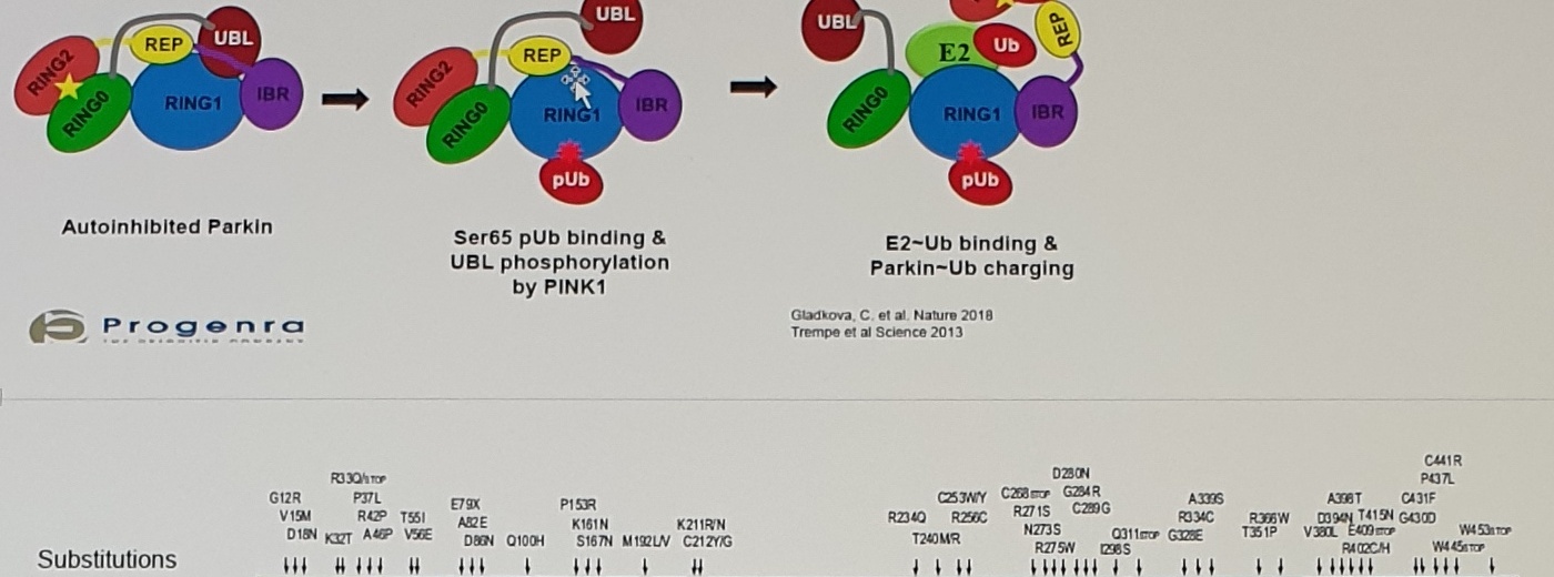
the tightly coiled conformation of parkin renders it inactive, as access to the catalytic RING2 residue is sterically blocked by RING0, while the E2 binding domain on RING1 is occluded by Ubl and REP.[6]
Activation
phosphorylation of serine Ser65 in Ubl by serine/threonine kinase, PINK1. Addition of a charged phosphate destabilises hydrophobic interactions between Ubl and neighbouring subregions, reducing autoinhibitory effects of this Nterminus domain.[13]
Activated status
disrupt these interdomain interactions and induce parkin to collapse along the RING1-RING0 interface.[12] The active site of RING2 is drawn towards E2-Ub bound to RING1, facilitating formation of the Ub-thioester intermediate.
note
-folded in half (?)
Parkin Activation State Diagram
Autoinhibited ParkinSer65 pUb binding & UBL phosphorylation by PINK1E2-Ub binding & Parkin~Ub chargingUBLREPRING2RING0RING1IBRpUbE2UbGladkova, C. et al. Nature 2018Trempe et al. Science 2013
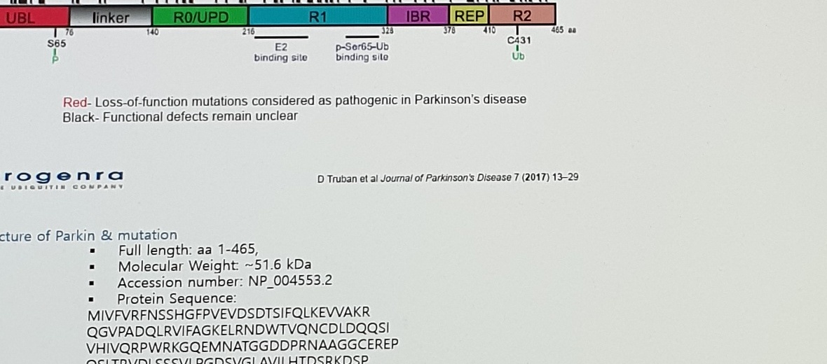
UBLlinkerR0/UPDR1IBRREPR2S65C431E2 binding sitep-Ser65-Ub binding siteUbRed - Loss-of-function mutations considered as pathogenic in Parkinson's diseaseBlack - Functional defects remain unclearD Truban et al Journal of Parkinson's Disease 7 (2017) 13-29
A member of RING-between-RING (RBR) family of E3 ligases,
Structure / Functional Notes Table
row
N term
UBL
RING0
RING1
IBR
RING2
C term
domain header
N term
UBL (ubiquitin-like domain)
RING0
RING1
IBR
RING2
C term
function
Autoinhibition at basal condition
linker
전체 parkin 이 반으로 접혀 있으면서, RING1 과 RING2 가 서로 마주보고 있다.
structural description
an innovative structure resembling
an N-terminal Ublike
a unique Parkin domain-UPD
binding site for E2 Ub-conjugating enzyme
Contains the catalytic cysteine residue (Cys431) that cleaves Ub off
zinc-finger / substrate note
zinc-finger domain
domain (Ubl) for specific substrate recognition,
E2 and transiently binds it to E3 via a thioester bond
mutation note
Deletion of UBL domain with/without the linker has little effect on Parkin activity (2).
deletion of RING0 (lack of occlusion of C431 as described) causes increased C431 reactivity and Parkin autoubiquitination (3).
275는 여기있네. A second class of mutations, including Thr240Arg, affect residues in and around the E2 binding site and alter autoinhibition of RING1 by REP.[3]
Cys431Phe and Gly430Asp mutations impair ligase activity at the catalytic site and significantly reduce parkin function.
2015 Fiesel variants
p.R33Q; p.P37L; p.R42P
p.C150G; p.K161N; p.W183A; p.K211N
p.T240R; p.R256C; p.Y267H; p.R275W
p.G328E; p.R334C
p.T415N; p.G430D; p.C431F; p.C431S; p.F463A
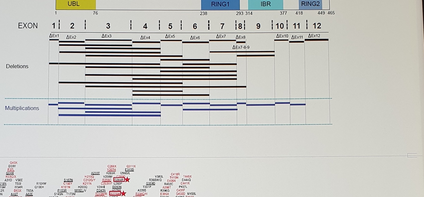
over 90% of them are located in exons 2, 3, 4 and 7 ("hot" exons), (2017 Likhachev)Mutations in hydrophobic residues of RING0:RING2 interface increase autoubiquitination (5).Firstly, those clustered around Zn-coordinating residues on RING and IBR might compromise structural integrity and impair catalysis.[12]
Substrates And Function Of Parkin
Substrates (& function) of Parkinfunctions in conjunction E2 enzymes UbcH7 (E2-640), UbcH8 (E2-644) and UbcH13/Uev1 (E2-664)https://resources.rndsystems.com/pdfs/datasheets/k-
Uncertain Spans
location
text/status
reason
exon/deletion map
substitution labels and deletion bars
Dense labels are asset-primary; selected mutation labels are transcribed but should not be treated as complete.
mutation domain map
red/black mutation labels
Asset preserves full visual; OCR is unreliable for many mutation names.
protein sequence
MIVF...FDV sequence
Long sequence is visible and OCR-supported but should be checked once before structured KB use.
Fiesel variant row
p.C150G, p.K161N, p.W183A, p.K211N, p.R275W, p.G430D, signs such as +, ++, -
Very small symbols and variant IDs; image asset should be primary evidence.